‘Three-parent babies’ explained

A 6-month-old baby boy may have opened the door to a new world of reproductive medicine. He is the first person born from a controversial new technique for preventing mitochondrial diseases by creating a “three-parent baby” — a child in which the vast majority of DNA comes from the mother and father and a small amount of DNA comes from a female donor.
Mitochondria are organelles inside cells that, among other tasks, generate energy. The organelles are passed from mother to child. Mutations in the 37 genes housed inside mitochondria can lead to fatal inherited diseases that affect organs that need lots of energy, such as the brain and muscles. There is no cure or effective treatment for many of the mitochondrial diseases.
Some of the mitochondria in the baby boy’s mother’s cells have a mutation that causes Leigh syndrome, a fatal neurological disorder. Most of her mitochondria function properly, so she doesn’t have the syndrome. But she can pass the disease on to her children: Two have died of the disease and she has had four miscarriages.
Her son’s birth in April caps nearly three decades of efforts to manipulate mitochondria and produce healthy eggs — initially to overcome fertility problems and now to avoid passing on disease. Although it takes three people to make these fertilized eggs, some researchers take issue with the moniker “three-parent baby.” Pioneering clinical embryologist Jacques Cohen calls the term erroneous. Mitochondrial DNA doesn’t contribute to a person’s traits, so a mitochondrial donor hardly constitutes a parent, he says.
Here’s a closer look at techniques that produce a baby that carries mitochondrial DNA from a donor:
Cytoplasmic transfer
In the late 1990s, Cohen and colleagues at Saint Barnabas Medical Center in Livingston, N.J., were looking for a way to help patients unable to have children by in vitro fertilization. The couples’ embryos did not develop normally for unknown reasons. Cohen and colleagues thought a dose of cytoplasm, the jellylike “guts” of a cell, from a donor egg might give the embryos a better shot at success.
“Cytoplasm is the most complicated fluid in the universe,” says Cohen. It contains mitochondria, other organelles, proteins and other molecules that do the work of the cell.
He extracted 10 to 15 percent of the cytoplasm from a donor egg and injected it along with a single sperm cell into a recipient egg. From 1996 to 2001, he performed the procedure 37 times, producing 17 babies for 13 couples.
At least two of eight children Cohen later tested carried detectable levels of donor mitochondria. Some of the other children may have had donor mitochondria at levels too low for his tests to detect at the time, he says. Cohen doesn’t know whether mitochondria or other cytoplasm components played a role in producing the children. He will soon publish a study reporting on the health of some of the children, who are now teenagers. Cohen’s group stopped performing the technique in 2001 because of regulatory issues.
Pronuclear transfer
The first “mitochondrial replacement” technique developed to stop mitochondrial diseases is called pronuclear transfer. It was first done in mouse embryos in 1983. Pronuclei are nuclei from the egg and sperm that are in the fertilized egg, called a zygote, but have not yet fused into a single nucleus.
In this technique, the mother’s egg and a donor egg are fertilized at the same time. The pronuclei are removed from the donor egg and discarded. Then the pronuclei are sucked out of the mother’s egg and transferred into the empty donor egg.
Pronuclear transfer has a couple of drawbacks. Some people object to it on ethical grounds because it is seen as destroying two embryos. Scientists worry because a bit of cytoplasm is usually transferred along with the pronuclei. That means that unacceptably high numbers of mitochondria — including disease-carrying ones — from the mother’s egg may be carried into the donor egg, says Shoukhrat Mitalipov, a mitochondrial biologist at Oregon Health & Science University in Portland.
In June, scientists reported that refinements in the technique produced embryos in which less than 2 percent of the mitochondria were carried from the mother’s egg into the donor egg (SN Online: 6/8/16). But an earlier study suggested that even 1 percent carryover could be dangerous because mutant mitochondria may replicate, eventually taking over the cell and crippling its energy production (SN: 6/25/16, p. 8).
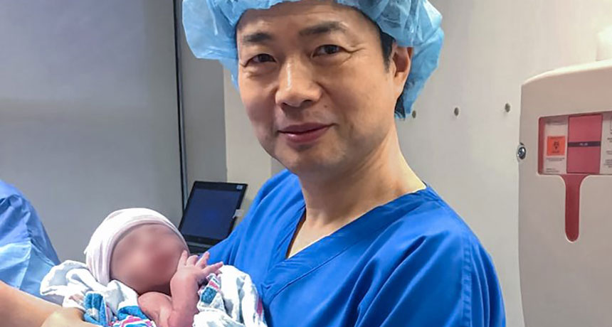
Fertility clinics in the United Kingdom are permitted to use this technique to make human babies, but none have reported doing so yet. New York fertility doctor John Zhang, the doctor involved in the baby boy’s case, tried the technique with colleagues at Sun-Yat Sen University of Medical Science in Guangzhou, China, more than a decade ago. Five embryos resulting from pronuclear transfer for a 30-year-old woman were implanted, and three grew into fetuses, but none survived to birth. Zhang published the results this year in Reproductive Biomedicine Online.
Spindle transfer
The technique used to produce the baby boy born in April is called spindle transfer. When a dividing cell divvies up its chromosomes, they are attached to protein fibers called microtubules or spindles. The transplant technique starts with two unfertilized egg cells, one from the donor and one from the mother. In both cells, the membrane surrounding the nucleus has broken down, but the cell has not yet completely divided.
The spindle and its attached chromosomes are removed from the mother’s egg and inserted into the donor egg, which has been emptied of its spindle and chromosomes. Then a sperm cell is injected to the resulting egg to fertilize it.
Mitalipov pioneered the spindle transfer technique, showing in 2009 that he could produce healthy monkeys (SN: 9/26/09, p. 8). The monkey experiments indicate that the technique has a lower level of carryover of mitochondria from the mother’s egg to the donor egg than pronuclear transfer, usually 1 percent or less, but Mitalipov would like to do even better. “This 1 percent is haunting us,” he says.
Spindle transfer has another possible downside: Chromosomes may fall off the spindle. That could result in an embryo with too few chromosomes — or too many if some are left in the egg from the donor or extras are carried over from the mother’s egg. Both cases usually result in abnormal development. Of five embryos Zhang performed spindle transfer on, only one developed normally to become the baby boy.
The infant reportedly has 1 percent of mitochondrial DNA from his mother. At 3 months old, he was healthy. Long-term consequences are unknown. Besides the risk of even trace levels of mitochondria ballooning, another study suggests that mismatches between the parents’ nuclear DNA and the donor mitochondrial DNA could affect aging (SN: 8/6/16, p. 8).